Watching laser-shocked stishovite grow from amorphous silica in nanoseconds – Dr. Wenge Yang & Dr. Wendy Mao
SEPTEMBER 4, 2015
Phase transitions have long been studied for centuries but little is known about the processes by which the atoms rearrange. New work from a team including HPSTAR scientists, Dr. Wenge Yang and Dr. Wendy Mao, performed in situ pump – probe x-ray diffraction measurements on shock-compressed fused silica, revealing the evolution of an amorphous to crystalline high pressure stishovite phase transition in nanoseconds. This is the first observation of shock-induced nucleation and growth of a high-pressure crystalline phase from an initially amorphous material. The study is published in recent Nature Communications (DOI: 10.1038/ncomms9191).
The high-pressure behavior of silica has been of long-standing scientific interest owing to its fundamental and technological importance and geophysical significance. The phase transition from crystalline quartz or amorphous silica to stishovite is one of the most studied high pressure transitions in materials science.
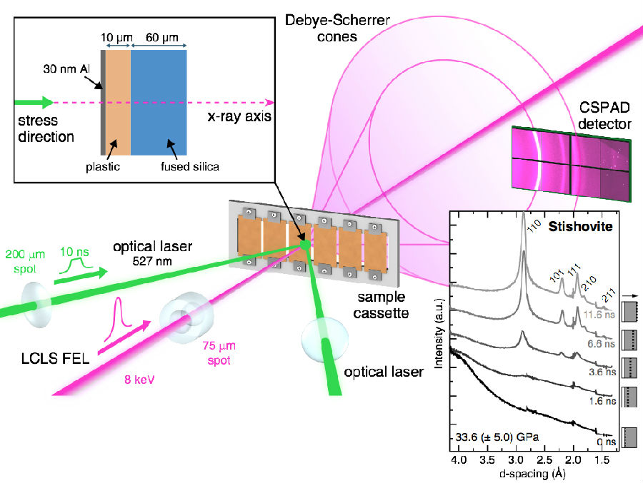
In the present work, the researchers utilized the intense, ultrashort pulses from a fourth generation synchrotron source, the X-ray free-electron laser (XFEL) at LCLS, to capture the evolution of the transitionin shock-compressed fused silica. Using LCLS, the researchers in situ observed the formation and growth of stishovite from shock-compressed amorphous silica over the course of several nanoseconds. Above 180, 000 atmospheric pressure (18 gigapasals), the nucleation of stishovite completed in less than 2 nanoseconds.

Caption: Experimental configuration of the XFEL probe and optical laser driver. The lattice response of the sample was captured in a Debye–Scherrer geometry. Inset: example of XRD resulting from azimuthal integration of CSPAD data for a suite of time delays under shock compression. A schematic of the target is shown on the right side for each time delay (white: plastic; grey: fused silica). A dashed line indicates the approximate location of the shock front; arrow is the shockpropagation direction.
Though the process from a disordered to ordered material was expected to be slow, the researchers found that the grains grew rapidly, and the growth trend supports a coalescence model rather than a diffusion-based mechanism.
This first demonstration of shock induced crystallization of an amorphous material via femtosecond diffraction will lead to a greater understanding of a number of important problems in shock physics, and their relation to geophysics. However, “our findings are only a starting place for exploring novel physics and new materials,” said Wendy Mao, one of the principal investigators on this project.
“We expect many more exciting discoveries as we explore shocked-induced phase-transition using XFELs,” said Wenge Yang, “which make it possible to capture detailed information during phase transitions on an ultrafast timescale.”
